Figure 2.

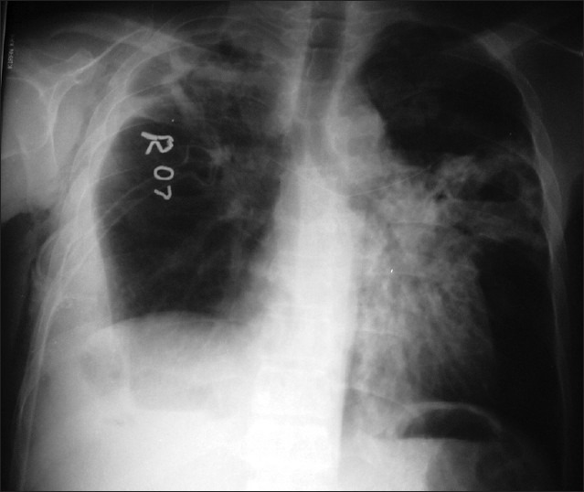
X-ray of the chest shows blunting of right costophrenic angle with intercostal tubein situalong with left mid-zone lung abscess and surrounding consolidation

X-ray of the chest shows blunting of right costophrenic angle with intercostal tubein situalong with left mid-zone lung abscess and surrounding consolidation