Figure 2.

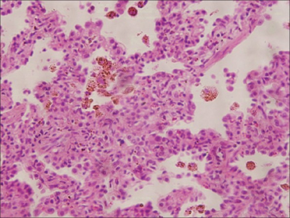
Photomicrograph shows type II pneumocyte hyperplasia, mild chronic inflammatory infiltrate in interalveolar septa and hemosiderin laden macrophages in alveolar spaces. No viral inclusions were identified. (H and E, ×100).

Photomicrograph shows type II pneumocyte hyperplasia, mild chronic inflammatory infiltrate in interalveolar septa and hemosiderin laden macrophages in alveolar spaces. No viral inclusions were identified. (H and E, ×100).