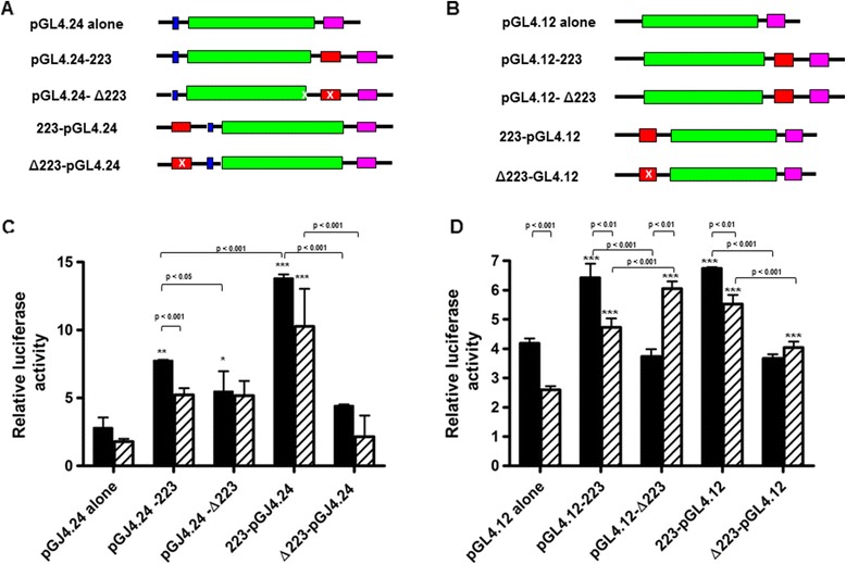
Figure 7.

Aca -DAF-16 drives expression of luciferase from Fragment 2.23. A. Fragment 2.23 (red) was cloned into different positions relative to reporter luciferase gene (green) in vector pGL4.24. Pink boxes represent the polyadenylation site of the vector, and blue boxes represent the minimal promoter. DBE deletion mutants are represented by an “x” in the red box. B. Fragment 2.23 was cloned into different positions relative to reporter luciferase gene in vector pGL4.12. C. Relative luciferase expression in NIH3T3 cells co-transfected with the pGL4.24 and pCMV4-DAF-16 constructs. Solid bars, no serum; striped bars, 20% serum. Results are normalized to empty vector control transfection. D. Relative luciferase expression in NIH3T3 cells co-transfected with the pGL4.12 and pCMV4-DAF-16 constructs. Significant differences between luciferase expression of Fragment 2.23 constructs and their appropriate plasmid alone control are denoted by asterisks (*p < 0.05, **p < 0.01, ***p < 0.001). Differences between other comparisons are shown with a bracket and p value. Differences within groups (serum or no serum) were determined by one-way ANOVA with Bonferroni’s Multiple Comparison post-test, and differences between serum and no serum were determined by unpaired t-test using GraphPad Prism.
