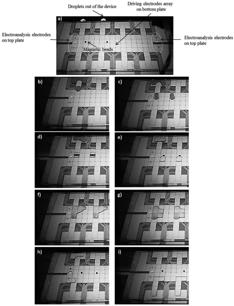
Figure 6.
Micro-graphs showing an example immunoassay on a two-plate EWOD DMF chip (reproduced with permission from [23]);(a–c) dispensing and transport of magnetic bead, antibody and blocking protein mixture; (d,e) trapping of magnetic beads and splitting of unused solvent mixture using a magnet; (f,g) washing and resuspension of the functionalized beads; (h,i) elution of the captured antibody target from the magnetic beads and electro analysis using the top plate measurement electrodes.

