
Fig. 3.
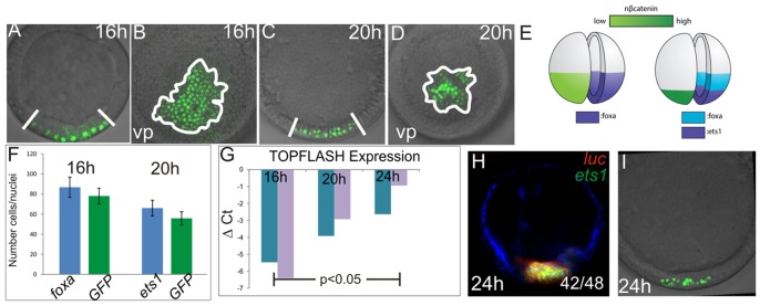
Spatiotemporal gradient of nβ-catenin in the vegetal pole. (A-D) Subcellular localization of β-catenin in 16-h- and 20-h-embryos assessed by confocal microscopy of GFP derived from the β-catenin:GFP fusion construct. 16-h-embryos are shown in A,B and 20-h-embryos in C,D. (A,C) Lateral views and (B,D) vegetal pole views. (E) Schematic of β-catenin nuclearization at 16 h and 20 h compared with expression of foxa and ets1 at these stages. (F) Comparison of number of GFP-positive nuclei and foxa- or ets1-positive cells at time points shown. Error bars are from one s.d. of ten individuals. (G) qPCR analysis of TOPFlash reporter expression in 16-h-, 20-h- and 24-h-embryos. Increased ΔCT (cycle threshold) values indicate increased reporter transcript abundance normalized to lamin2β receptor. (H) FISH showing co-expression of luciferase driven by the TOPFlash reporter and the mesoderm marker ets1 at 24 h. Co-expression of ets1 and luciferase was detected in 42 of 48 embryos (expression in the presumptive endoderm was detected in five embryos). (I) Subcellular localization of the β-catenin:GFP fusion construct in 24-h-embryos for comparison with (H) at the same stage.