Fig. 3.
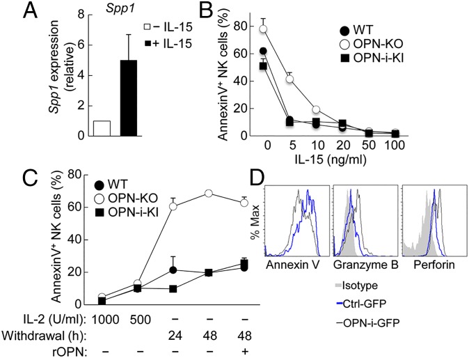
Impaired IL-15 responses of OPN-KO NK cells in vitro. (A) Quantitative RT-PCR analysis of Spp1 mRNA expressed by B6 splenic NK cells treated with (+) or without (–) IL-15 (100 ng/mL) for 24 h. Results (normalized as in Fig. 1A) are presented relative to that of (–), set as 1. (B and C) Percent of annexin V+ NK cells (from the indicated mice) incubated with increasing concentrations of IL-15 for 24 h (B) and with or without IL-2 at the indicated time points (C), as well as with (+) or without (–) addition of recombinant OPN (C), assessed by flow cytometry. Values represent the average of three independent experiments with error bars of mean ± SEM. (D) Lentivirally transduced OPN-KO HSCs were transferred into Rag2−/−γC−/− hosts as in Fig. 2B. Expression of annexin V, intracellular granzyme B, and perforin was analyzed by flow cytometry in splenic NK cells 8 wk after reconstitution followed by incubation with IL-15 (100 ng/mL) for 24 h.

