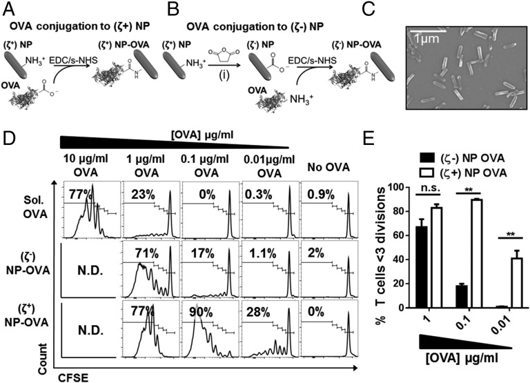
Fig. 1.
CD4+ OT-II T-cell proliferation in response to BMDCs treated with ovalbumin functionalized PRINT particles. (A) Model antigen OVA was covalently linked to cationic (ζ+) monodisperse PRINT 80 × 80 × 320 nm particles by using EDC/s-NHS carbodiimide chemistry yielding (ζ+)NP-OVA. (B) Amine groups in (ζ+)NP were converted to carboxylic groups by using succinic anhydride (i) to yield anionic (ζ−)NP that were covalently linked to OVA by using the same chemistry as in A. (C) Representative scanning EM micrograph of functionalized NPs. (D) Representative CFSE dilution plots of OVA-specific CD4+VB5.1+ OT-II T-cell division after 72 h coculture with BMDC treated with equivalent does of OVA protein. Soluble OVA (Upper), (ζ−) NP-OVA (Middle), and (ζ+) NP-OVA (Lower). Number represents frequency of OVA-specific CD4+ T cells that underwent >3 divisions. N.D., not done. (E) Combined data from three experiments described in A by using independently synthesized NP-OVA batches. **P < 0.001 two-way ANOVA with Sidak’s multiple comparisons test. Data graphed as mean ± SEM.

