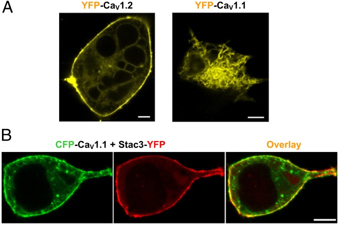
Fig. 1.
CaV1.1 is retained in the ER of tsA201 cells but traffics to the surface when Stac3 is present. (A) Subcellular distribution of YFP-labeled CaV1.2 and CaV1.1 in tsA201 cells. (Scale bars, 5 μm.) (B) Fluorescence images demonstrating that CFP-CaV1.1 (green) and Stac3-YFP (red) traffic to the surface after coexpression in tsA201 cells. (Scale bar, 2 μm.) The auxiliary β1a- and α2δ-subunits were also present here and in all other tsA201 cell experiments, unless otherwise mentioned.

