Fig. 4.
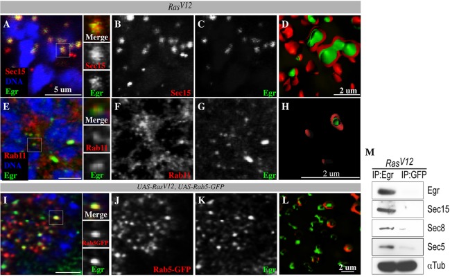
Sec15 interacts with Eiger/TNF. (A-D) RasV12 cells counterstained with anti-Sec15 (red) and anti-Eiger (green). Sec15 and Eiger colocalize (A; side panels represent magnified view from white box). B and C are single channels for Sec15 and Eiger from A. (D) Cross-section images of isosurface-rendered confocal projection images showing Eiger (green) inside Sec15-coated (red) vesicles. (E-H) RasV12 cells counterstained with anti-Rab11 (red) and anti-Eiger (green). Rab11 and Eiger colocalize (E; side panels represent magnified view from white box). F and G are single channels for Rab11 and Eiger from E. (H) Cross-section of isosurface-rendered confocal projection showing Eiger (green) inside Rab11-coated (red) vesicles. (I-L) RasV12 cells co-expressing Rab5-GFP (red) counterstained with anti-Eiger (green). Rab5-GFP and Eiger colocalize (I; side panels represent magnified view from white box). J and K are single channels for Rab5-GFP and Eiger, respectively. (L) Cross-section of isosurface-rendered confocal projections showing Eiger (green) inside Rab5-GFP-coated (red) vesicles. (M) Anti-Eiger or anti-GFP (control) immunoprecipitation experiments with lysate derived from tissues bearing RasV12 clones. Immunoprecipitates (IP) were blotted with antibodies against Sec15, Sec8, Sec5 and α-Tubulin (loading control).

