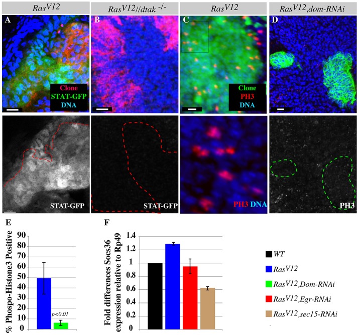
Fig. 6.
Eiger/TNF exocytosis activates JAK-STAT signaling through JNK. (A) The JAK-STAT activity GFP-reporter line shows pathway activation in RasV12 clones. The lower panel shows STAT-GFP alone. (B) dTAK1-rescued RasV12 clones (red) induced in dtak1 hemizygous mutant discs do not show JAK-STAT GFP (green) induction. Lower panel shows STAT-GFP alone. The red outlines show clone boundaries in A and B. (C,D) RasV12 or RasV12, dom-RNAi double mutant clones stained with DAPI to mark nuclei and with antibody against the mitotic marker, phospho-histone-3 (PH3). The majority of RasV12 clones are phospho-histone-3 positive (C). The lower panel of C represents a magnified view of DAPI and phospho-histone-3 from the boxed area in the upper panel. RasV12, dom-RNAi clones (D) show reduced phospho-histone-3 staining. The lower panel shows the phospho-histone-3 channel alone. The green outlines show clone boundaries in D. Scale bars: 5 μm. (E) Quantification of C and D. (F) RT-PCR experiment measuring Socs36E levels relative to expression levels of the housekeeping gene Rp49. Normalized fold differences in Socs36E expression levels between RasV12 and wild-type (WT) or between RasV12, eiger-RNAi (eiger RNAi-knockdown specifically in RasV12 clones) and RasV12 or between RasV12, sec15 double mutant and RasV12 clones are shown. Experiments were performed in triplicate and standard deviations were derived from the coefficient variations of experimental and control samples.

