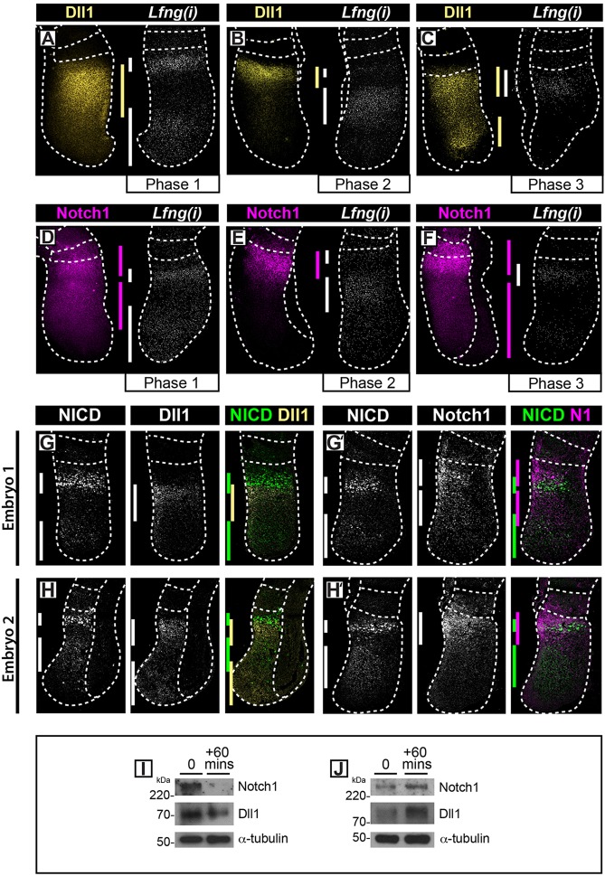
Fig. 5.
Oscillations of Dll1 and Notch1 proteins in mouse PSM. (A-F) Following FISH to detect Lfng(i) in one half of a set of E10.5 tails, immunohistochemistry was performed on the contralateral half of the explants to detect Dll1 (A-C) or Notch1 (D-F) protein. (G-H′) Double immunohistochemistry on sections of individual E10.5 tails to detect PSM expression of NICD and Dll1 (G,H), or NICD and Notch1 (G′,H′) (n=15). The dotted lines demarcate the positions of the most recently formed somite(s), outer edges of the PSM, adjacent neural tissue (C,E) or hind gut (H). (I,J) Western blot analysis for Dll1 and Notch1 or α-tubulin on the caudal half of individual PSM explants following fix and culture.

