Fig. 2.
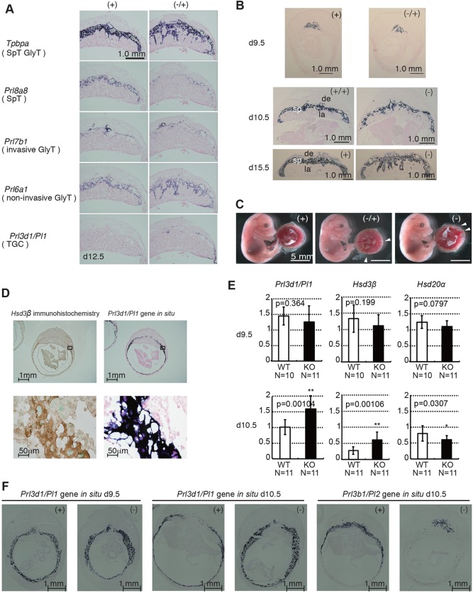
Abnormal differentiation of Sirh7/Ldoc1 KO placentas. (A) Expression patterns of trophoblast lineage-restricted markers in the d12.5 placenta by RNA in situ hybridization. Tpbpa, Prl8a8, Prl7b1, Prl6a1 and Prl3d1 identify cells specific for spongiotrophoblast cells (SpT) and GlyT, SpT, invasive GlyT, non-invasive GlyT, and TGCs, respectively. Invasion of the Prl6a1-positive GlyTs took place in the labyrinth layer in only Sirh7/Ldoc1 KO. There were fewer Prl8a8-positive SpTs in Sirh7/Ldoc1 KO on d12.5. (B) Irregular spongiotrophoblast layer formation on d9.5, d10.5 and d15.5 (see A, Tpbpa on d12.5). (C) Fetuses and placentas on d15.5. The arrowheads indicate the regions where the labyrinth layers almost reached the maternal decidua. Despite the placental abnormality, Sirh7/Ldoc1 KO fetuses looked normal (see also supplementary material Fig. S4). (D) Immunohistochemistry using an anti-Hsd3β antibody (left) and in situ hybridization for Prl3d1 on d10.5 wild-type placenta (right). Higher magnifications of the boxed region of upper panels are shown in lower panels, respectively. (E) Relative expression levels of Prl3d1, Hsd3b and Hsd20a on d9.5 and d10.5 in each wild-type (+ or +/+) and KO (− or −/−) placenta (the expression level of β-actin was arbitrarily assigned a value of 1). White and black bars represent wild-type and KO placentas, respectively. Data are average±s.d. (**P<0.01, *P<0.005). (F) Expression patterns of the TGC markers Prl3d1 (d9.5 and d10.5) and Prl3b1 (d10.5) in wild-type and Sirh7/Ldoc1 KO placenta by RNA in situ hybridization.

