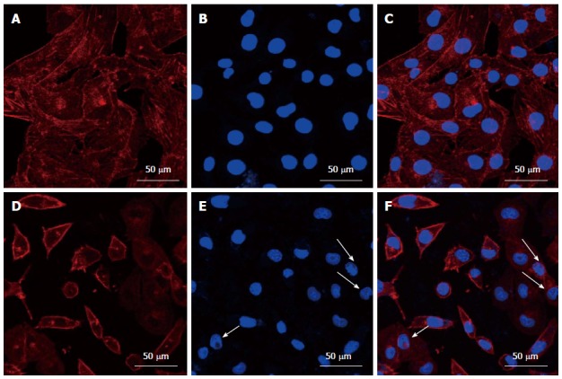
Figure 3.

Morphology of the cytoskeleton and nuclei in HepG2 cells before (A-C) and after (D-F) treatment with cinobufacini. The cytoskeleton and nuclei were stained with rhodamine-phalloidin and DAPI. (C) and (F) were merged images of (A, B) and (D, E). White arrow: Fragmented nuclei.
