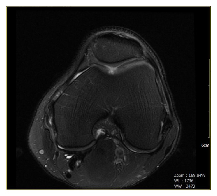
Figure 1.

Preoperative radiograph. Multiplanar, multisequential images were obtained through the left knee on a 1.5 Tesla MRI scanner. Mild patellofemoral joint osteoarthritis. A focal region of grades III-IV chondromalacia overlying the central femoral trochlear groove.
