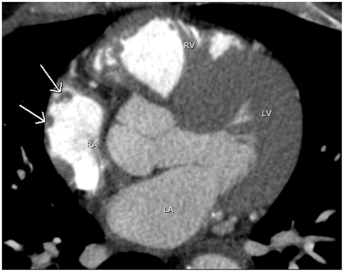
Fig. 1.
A 44-year-old man with atrial fibrillation. A pulmonary vein CT for pre-ablation mapping shows multiple small filling defects abutting the lateral wall of the right atrium (arrows), compatible with prominent pectinate muscles. Note contrast opacification in the right heart due to poor timing. LA, left atrium; LV, left ventricle; RA, right atrium; RV, right ventricle.

