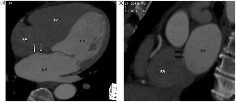
Fig. 10.
A 74-year-old woman with left ventricular thrombus seen on echocardiogram. Cardiac CT was performed to rule out left ventricular thrombus. The normal thinning of interatrial septum at the fossa ovalis is seen adjacent to the left atrial pouch, corresponding to the unfused septum primum and secundum on four chamber (double white arrows in a) and sagittal oblique views (double white arrows in b). This can be mistaken for an atrial septal defect.

