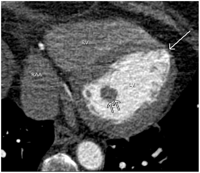
Fig. 16.
A 66-year-old woman with atrial fibrillation. A pulmonary vein CT for pre-ablation mapping shows a physiologic apical thinning (arrow). Physiologic thinning of the LV apex is occasionally mistaken as a sequlae of myocardial infarction. Note the prominent posterior papillary muscle (double arrows). RAA, right atrial appendage.

