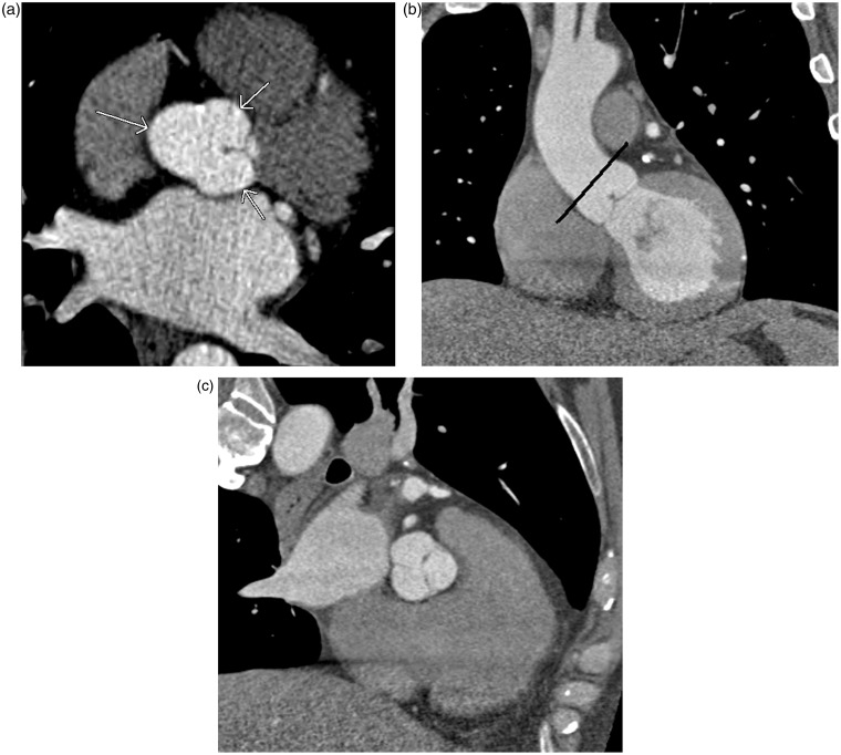
Fig. 18.
A 56-year-old man with atrial fibrillation. A pulmonary vein CT for pre-ablation mapping shows distortion of the Sinus of Valsalva of the aorta which can simulate the sinus of Valsalva aneurysm (arrows in a). Images perpendicular through the aortic valve (black line in b) will clarify this normal anatomy (c).

