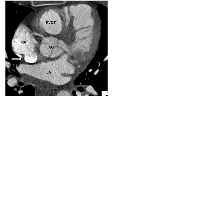
Fig. 2.
A 62-year-old man with chest pain and prior myocardial infarction. Multiplanar images of a coronary CTA for coronary artery evaluation show the normal crista terminalis identified as a smooth soft tissue ridge along the posterolateral wall of right atrium on axial (white arrow in a), coronal and sagittal planes (black arrow in b, c). AO, aorta; RVOT, right ventricular outflow tract; SVC, superior vena cava.

