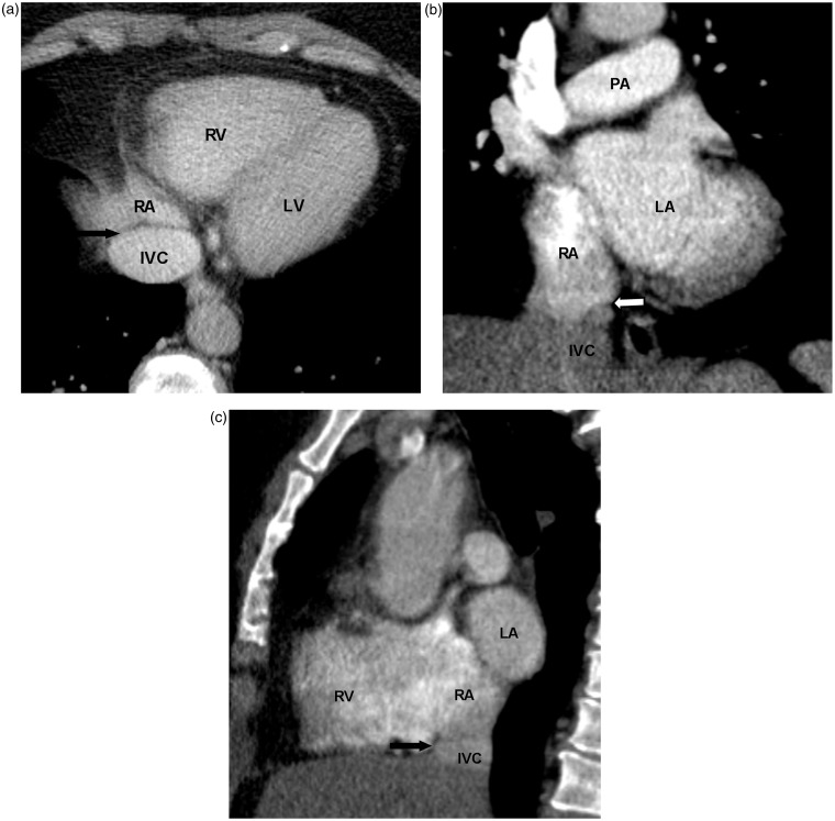
Fig. 3.
A 61-year-old woman with head and neck cancer. Chest CT with contrast was performed for restaging. Normal Eustachian valve is seen as a paper-thin structure at the junction of IVC and RA on axial (black arrow in a), coronal (white arrow in b), and sagittal planes (black arrow in c). IVC, inferior vena cava; PA, pulmonary artery.

