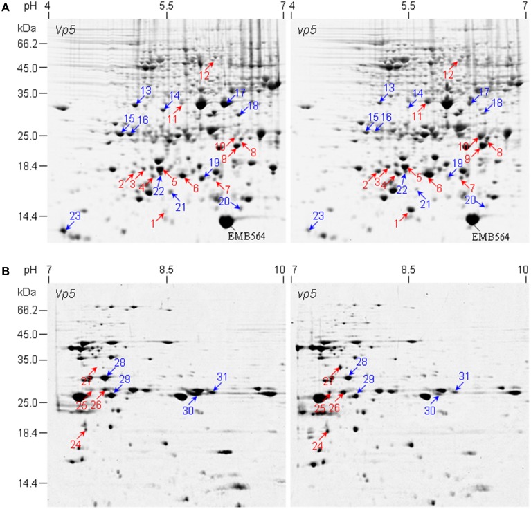
Figure 1.
2-DE comparison of embryo protein profiles between maize mutant vp5 and wild type Vp5. (A,B) are representative 2-DE gels of pH 4–7 and pH 7–10, respectively. Differentially accumulated protein spots between vp5 and Vp5 embryos are indicated by arrows. Red arrow, increased abundance in vp5; blue arrow, increased abundance in Vp5. Embryo protein was extracted and resolved in 2-DE with IEF in the first dimension and 13.5% SDS-PAGE gel in the second dimension. Gels were stained with colloidal CBB G250. The identified proteins were listed in Tables 1, 2.

