Figure 5.
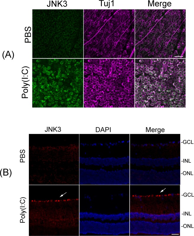
Upregulation of JNK3 protein in Tuj1-positive RGCs. (A) Whole retinas isolated from PBS- or Poly(I:C)-treated eyes (at 48 hours) were immunostained with antibodies against JNK3 and Tuj1. Images were obtained at ×20 magnification. Scale bars: 50 μm. Results presented indicate that Poly(I:C)-mediated upregulation of JNK3 was localized in Tuj1-positive RGCs, but not in other cell types. (B) Localization of JNK3 protein in RGCs. Retinal cross sections prepared from Poly(I:C)- or PBS-treated eyes (at 48 hours) were immunostained by using antibodies against JNK3. Retinal cross sections were stained with DAPI to identify the nuclei. Representative immunohistochemistry results indicate that Poly(I:C)-mediated upregulation of JNK3 protein is localized in RGCs (white arrows). Images were obtained at ×40 magnification.

