
Figure 6. Propagation of H3K9me3 by stably tethered SUV39H1 to nucleosomes in its spatial proximity.
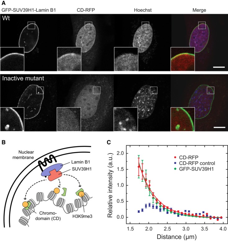
- Establishment of H3K9me3 domains by GFP-SUV39H1 recruited to the nuclear lamina via GBP-Lamin B1 in iMEF Suv39h dn cells. H3K9me3 was detected via CD-RFP and appeared in a confined region adjacent to the nuclear lamina (see inset). When the inactive mutant SUV39H1-H324L-GFP was recruited, no enrichment of CD-RFP was observed. Scale bar, 10 μm.
- Cartoon model depicting the experimental setup in panel A.
- Averaged radial fluorescence intensity profiles from the lamina to the center of the nucleus measured for the experiments described in panel A. The profile of CD-RFP reflects the H3K9me3 levels and was measured in cells transfected with GFP-SUV39H1 (red) or the inactive SUV39H1-H324L-GFP mutant (blue, control). The recruitment of GFP-SUV39H1 (green) resulted in a lamina-confined enrichment with a width of 0.40 ± 0.02 μm as determined by fitting the data to an exponential decay curve. While wild-type SUV39H1 methylated the surrounding chromatin within a confined area of 0.47 ± 0.03 μm width (red), SUV39H1-H324L (blue) did not increase the methylation (blue) in this region. Error bars correspond to SEM.