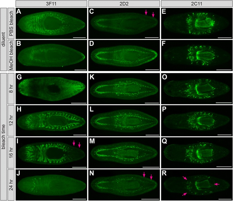
Figure 5.

Bleaching affects mAb labeling. (A-B) Planarians labeled with mAb 3F11 after hydrogen peroxide bleaching in either PBS (A) or methanol (B). Labeling is completely abolished by methanol bleaching. (C-D) mAb 2D2 labeling of the CNS is more robust in samples bleached in methanol, particularly in posterior regions (arrows indicated reduced signal in C). (E-F) mAb 2C11 labeling is unaffected by bleaching diluent. (G-J) Planarians labeled with mAb 3F11 after bleaching for the times indicated at left. Arrows (I) indicate signal loss in tail branches. (K-N) Planarians labeled with mAb 2D2. Arrows (N) indicate signal loss in tail branches. (O-R) Planarians labeled with mAb 2C11. Arrows (R) indicate decreased signal both within and around the pharynx. Planarians were relaxed in magnesium chloride, treated with 2% HCl (3F11) or 7.5% NAc (others), fixed in formaldehyde/Triton X-100, and bleached in 6% H2O2/PBS (3F11) or 6% H2O2/methanol (others). Samples were bleached for 12 hr in A-F. Scale bars: 500 μm.
