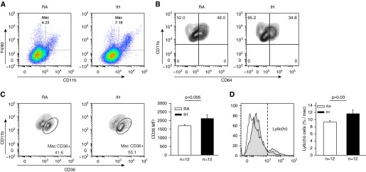
Figure 1.

Changes in macrophage populations in the aortic wall of mice exposed to intermittent hypoxia (IH) or room air (RA) during sleep for 6 weeks. (A) Representative example of fluorescence-activated cell sorter analysis of macrophages (CD11b and F4/80 positively labeled cells) and the increase in such population with IH. (B) Representative example of fluorescence-activated cell sorter analysis for CD64+ cells (resident macrophages) illustrating the reduction in resident macrophages after IH exposures. (C) IH is associated with significantly increased expression of CD36 in CD11b and F4/80 positively labeled cells indicating metabolic activation. (D) IH induced significant increases in Ly6C(hi) cell expression, indicating shifts toward proinflammatory macrophages recruited from the bone marrow. MFI = mean fluorescence intensity.
