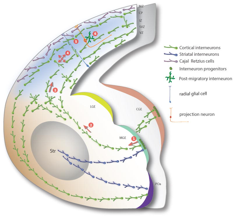
Figure 1. Patterns of interneuron migration in the developing telencephalon.
This schema shows rostral and caudal hemi-section through the mouse telencephalon at mid-embryonic (E15) stage. The major decision-making steps (1-6) involved in the migration of cortical interneurons derived from the subpallium are illustrated. Interneurons derived from MGE (green), POa (purple), or CGE (orange) exit the proliferative zones and initiate their migration towards the developing neocortex and striatum. Cortical interneurons traverse around the developing striatum, transit across the cortico-subpallial boundary, and course tangentially into the cortex, whereas striatal interneurons ventrolaterally migrate into the developing striatum. Cortical interneurons transit the neocortex mainly through the MZ, SP, IZ/SVZ migratory streams. Once in the neocortex, tangentially migrating interneurons undergo multi-modal local migration as they reach and settle in specific areal and laminar locations within the emerging CP, prior to forming functional synaptic contacts with appropriate projection neuron partners. Multiple decision-making steps are involved in this process. These include: (1) exit from the proliferative zone and initiation of migration in the subpallium, (2) selection of migratory route towards the dorsal cortex, (3) choice of migratory streams within the neocortex, (4) local orientation of migration within the cerebral wall, (5) identification of the final areal and laminar location, and (6) termination of migration at the appropriate cortical layer. Arrows indicate net directionality of movement. LGE, lateral ganglionic eminence; MGE, medial ganglionic eminence; POa, preoptic area; Str, striatum; MZ, marginal zone; CP, cortical plate; IZ, intermediate zone; SVZ, subventricular zone; VZ, ventricular zone.

