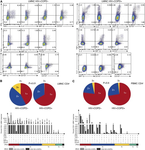
Figure 3.
Loss of multifunctional HIV-specific CD4+ T-cell memory in the lung mucosa in HIV-associated chronic obstructive pulmonary disease (COPD). (A) Representative multifunctional flow cytometric plots of lung mononuclear cells (LMNC) from HIV+COPD− (left) versus HIV+COPD+ (right), following restimulation with Pol peptides showing multifunctional subset frequencies of CD4+ T cells producing IFN-γ, tumor necrosis factor (TNF)-α, IL-2, MIP-1β, and CD107 during chronic HIV infection. (B) Individual pie charts of LMNC CD4+ T cell reflecting the percentage of Pol-specific CD4+ T cells from HIV+COPD− (left pie chart) versus HIV+COPD+ (right pie chart) patients producing one, two, three, or four functional responses. (C) Individual pie charts of peripheral blood mononuclear cells (PBMC) CD4+ T cells, reflecting the same cohort responses as in B. Using Boolean analysis, the percentage of total for individual multifunctional subset responses for HIV+COPD− (gray bars) and HIV+COPD+ (black bars) are shown in the bar graph for each of the multifunctional subsets (B and C). Significant differences when comparing mean frequencies of Pol-specific single and multifunctional responses are indicated by *P < 0.05, **P < 0.01, and ***P < 0.001. All P values were determined by the Kruskal-Wallis one-way analysis of variance or Wilcoxon signed rank test.

