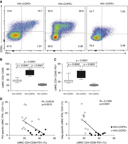
Figure 4.
HIV+COPD+ patients have a significantly higher surface expression of CD95+ and/or programed death (PD)-1+ on lung mucosal CD4+ T cells. (A) Representative flow cytometric plots of the lung mononuclear cells (LMNC) CD4+CD95+PD-1+ for HIV+COPD− (left) versus HIV+COPD+ (middle), and HIV−COPD+ (right) patient. (B) Pooled data showing cumulative frequencies of LMNC CD4+CD95+ for 11 HIV+COPD−, 12 HIV+COPD+, and 7 HIV−COPD+ patients. (C) Pooled data showing cumulative frequencies of LMNC CD4+CD95+PD-1+ for 11 HIV+COPD− patients (white bars), 12 HIV+COPD+ (black bars), and 7 HIV−COPD+ (gray bars) patients. Values represent mean ± SEM, and P values were calculated using the Mann-Whitney t test. (D) Scatterplot analysis of LMNC Pol- and Gag-specific CD4+IFNγ+ responses and CD4+CD95+PD-1+ frequencies. Values are for 12 HIV+COPD+ (black circles) and 11 HIV+COPD− (white circles) individuals in the cohort. Analysis was performed using the Spearman rho test.

