Figure 2.
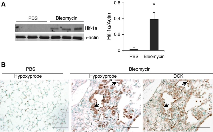
Hypoxia is present in the lungs of mice exposed to bleomycin and colocalized with deoxycytidine kinase (DCK). (A) Western blot analysis and quantitative densitometry of the protein expressions of hypoxia-inducible factor 1α (Hif-1a) in whole lung lysates at Day 33 after phosphate-buffered saline (PBS) or bleomycin exposure. Different lanes represent samples collected from distinct mice. α-Actin was used as protein loading control. *P < 0.01 versus PBS. (B) Hypoxyprobe, to identify hypoxic cells, was injected intraperitoneally into PBS (left) or bleomycin (middle) treated mice. Lungs were collected after 90 minutes and immunohistochemistry was carried-out using antihypoxyprobe antibodies to localize the hypoxic cells (left and middle). An adjacent slide was immunostained for DCK to visualize DCK localization (right). Scale bar = 100 μm. Arrows represent hyperplastic alveolar epithelial cells.

