Figure 8.
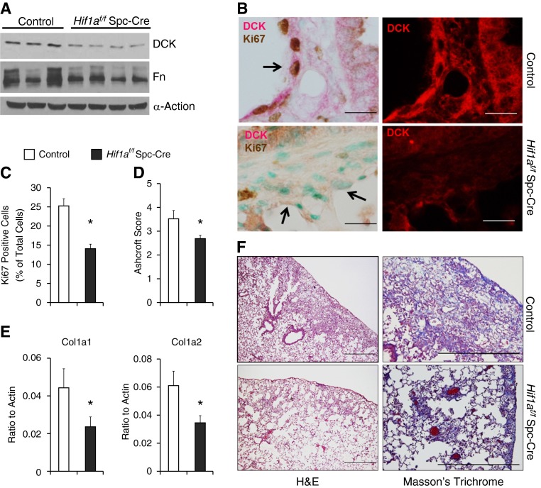
Tissue-specific function of hypoxia-inducible factor 1α (Hif-1a) in pulmonary fibrosis. Hif1af/f Spc-Cre mice and corresponding age-, weight-, and sex-matched littermate control animals (Cre-expressing mice) were exposed to bleomycin. (A) The protein levels of deoxycytidine kinase (DCK) and fibronectin (Fn) in the lungs of mice exposed to bleomycin were determined using Western blot. α-Actin was used as an internal control. (B) Dual immunohistochemistry was performed in the lungs of mice exposed to bleomycin to visualize the colocalization of DCK with Ki-67. Scale bar = 100 μm. Arrows represent hyperplastic alveolar epithelial cells. (C) The percentage of Ki-67–positive cells were calculated. (D) Ashcroft scores were blindly assigned to evaluate levels of pulmonary fibrosis. (E) Transcript levels of Col1a1 and Col1a2 were determined using real-time polymerase chain reaction. n = 3 for control, n = 4 for Hif1af/f Spc-Cre. (F) Lung sections were stained with hematoxylin and eosin (H&E; left) or Masson trichrome (right) to visualize histology and levels of collagen deposition, respectively. Scale bar = 500 μm. n = 6 for control, n = 15 for Hif1af/f Spc-Cre. *P < 0.05 versus control.

