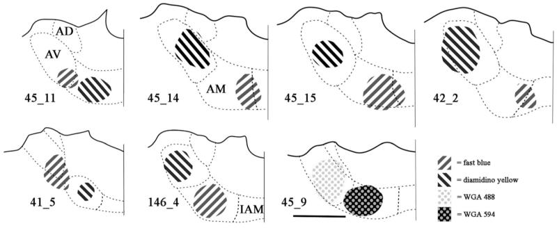
Fig. 1.
Line drawings constructed from photomicrographs of the injection sites located in the anteroventral (AV) and anteromedial thalamic (AM) nuclei from selected cases. Fast blue is indicated by diagonal stripes to the right. Diamidino yellow is indicated by diagonals to the left. WGA488 is indicated by grey dots on a white background. WGA594 is indicated by pale dots on a black background. Scale bars = 1 mm. All sections are in the coronal plane, though it should be noted that the injections were spherical in shape remaining within the target nuclei. For case 41_5, the injection into AM is located approximately 280 μm posterior to the injection in AV. Abbreviations as Table 1.

