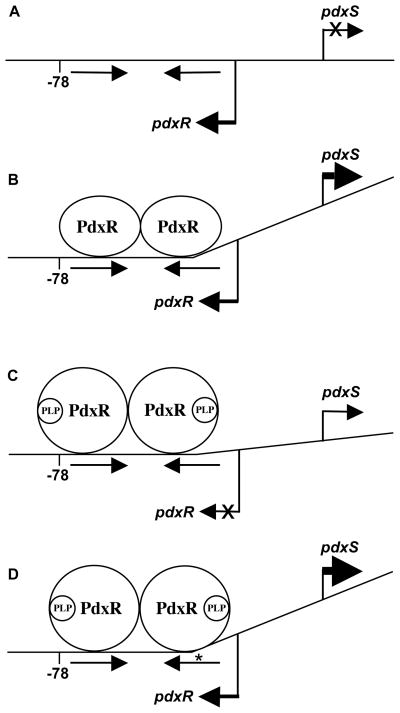
Fig. 8. A model of PdxR interaction with the wild-type and mutant pdxRS regulatory regions.
(A) wild-type DNA, no PdxR; (B) wild-type DNA plus PdxR; (C) wild-type DNA plus PdxR and PLP; (D) p2- or p4-containing DNA plus PdxR and PLP. The model (not to scale) assumes that a PdxR dimer binds to the dyad-symmetry element (indicated by straight arrows) and includes hypothetical PLP-dependent bending induced by PdxR. Bent arrows show the start points and directions of transcription. An asterisk indicates the location of the adjacent p2 and p4 mutations. The position -78 (with respect to pdxS), protected by the PLP-bound form of PdxR on wild-type DNA, is shown.

