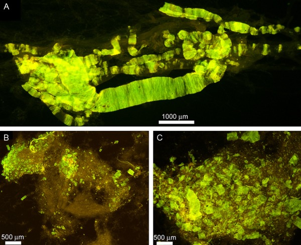
Figure 3.

Whole mounts of cerebral vessels exhibiting amyloid angiopathy in the Semagacestat-treated patient. (A) Cortical vessels stained by thioflavine-S. To isolate the vessels small cubes of cerebral cortex, approximately 0.5 cm, were stirred in 50 mM Tris-HCl buffer pH 7.5 containing 5% SDS. After 72 h the only remaining structures are the insoluble extracellular matrix of the vasculature and associated amyloid deposits. Magnification 100X. (B and C) Leptomeninges showing abundant clusters of vascular amyloid deposits stained by thioflavine-S. Magnifications: (B) 25X and (C) 100X.
