Figure 1.

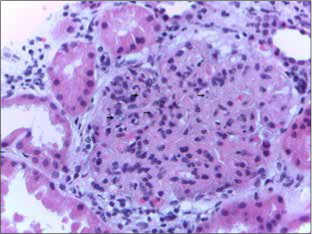
Glomerulus showing lobulation of the capillary tuft, mesangial hypercellularity, and thickening of the capillary walls. Leishmania amastigotes are seen within capillary lumens (arrows). HE ×400.

Glomerulus showing lobulation of the capillary tuft, mesangial hypercellularity, and thickening of the capillary walls. Leishmania amastigotes are seen within capillary lumens (arrows). HE ×400.