
Figure 5.
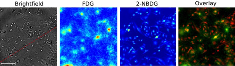
Radioluminescence microscopy results of FDG and 2-NBDG. Reprinted with permission from Pratx et al. 2012 [42]. Scale bar is 100 µm.
Official websites use .gov
A
.gov website belongs to an official
government organization in the United States.
Secure .gov websites use HTTPS
A lock (
) or https:// means you've safely
connected to the .gov website. Share sensitive
information only on official, secure websites.
Radioluminescence microscopy results of FDG and 2-NBDG. Reprinted with permission from Pratx et al. 2012 [42]. Scale bar is 100 µm.