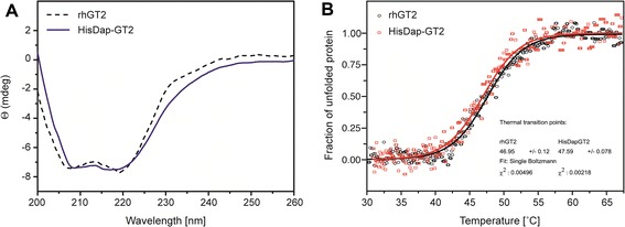
Figure 4.

CD spectroscopy and thermal stability measurements of HisDapGalNAcT2 in comparison to rhGalNAcT2. (A) Far UV (195–260 nm) spectra of HisDapGalNAcT2 (solid line) and rhGalNAcT2 (dashed line) indicating the folded status of the proteins at 21°C in 20 mM sodium phosphate buffer at pH 7.3 CD data are reported as molar ellipticity (Θ) [24]. (B) Thermal unfolding profiles of HisDapGalNAcT2 (squares) and rhGalNAcT2 (circles) were monitored at 208 nm.
