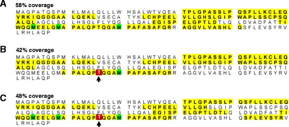
Figure 7.

ESI-MS/MS analysis of non-glycosylated and glycosylated Filgrastim™ (G-CSF). (A) Amino acid sequence of the substrate Filgrastim™ showing the tryptic digest peptides detected by ESI-MS/MS (yellow highlight, 58% coverage) and post-translationally modified amino acids (e.g. oxidation, green). G-CSF peptides detected by ESI-MS/MS after treatment with glycosyltransferases (B) HisDapGalNAcT2 (42% coverage) or (C) of rhGalNAcT2 (48% coverage), showing the glycosylation site at Thr166 (red box and black arrow).
