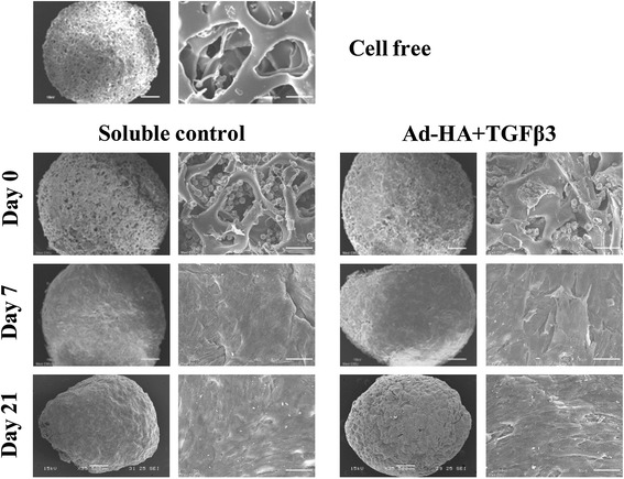
Figure 6.

Scanning electron microscopic examination of chondrocyte-seeded gelatin scaffolds. A Scanning electron microscope was used on the surface of the cell-free gelatin scaffolds, chondrocyte-cultured HA + TGF-β3 adsorbed scaffolds, and soluble control scaffolds on days 0, 7 and 21 of cultivation. Images are at 35x and 500x insets.
