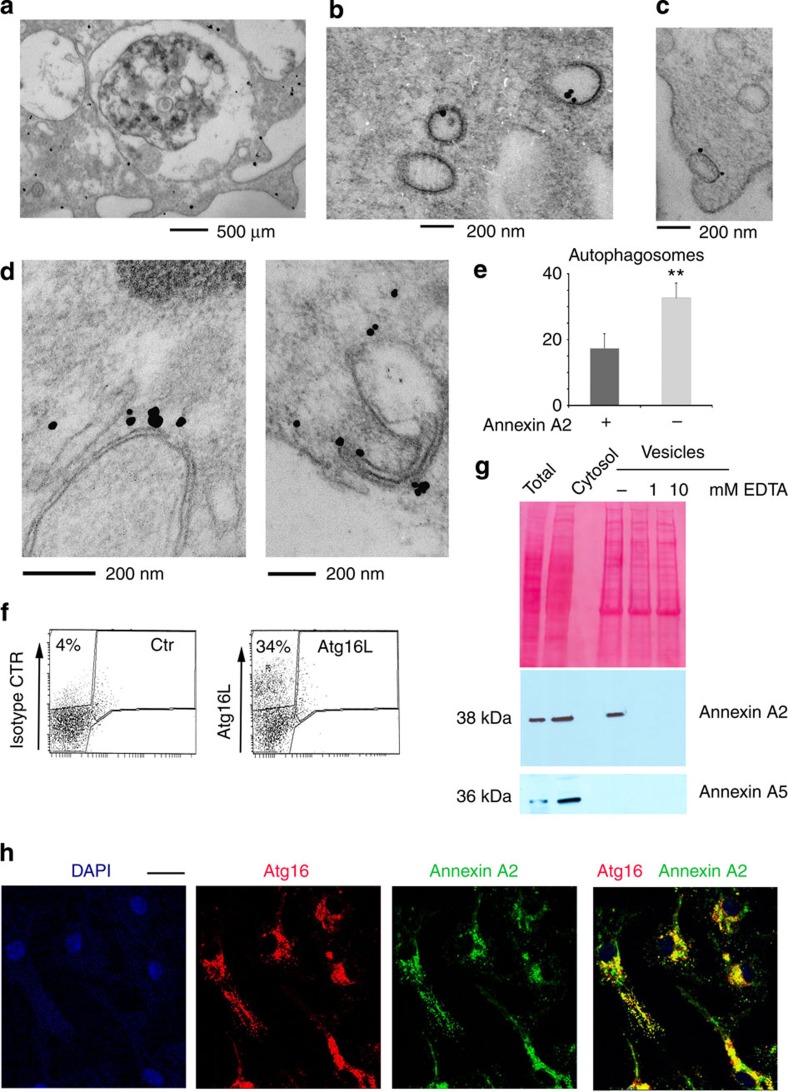
Figure 1. DC phagophore precursors and autophagosome are annexin A2-positive.
(a–c) Immunogold staining of dendritic cells with annexin A2 antibody identifies positive staining in the (a) cytosol, (a) plasma membrane, (a) multivesicular late endosomes (MVB) and (b,c) vesicular structures: n≥50 cells. (d) Immunogold labelling of phagophore structures with annexin A2 antibody; n≥50 cells. (e) Quantification of annexin A2-positive and -negative autophagosomes in primary dendritic cells; the mean and s.e. were calculated from n≥50 cells: P<0.01 (unpaired two-tailed Student’s t-test). (f) FACS analysis of total DC vesicles prepared by cytosol ultracentrifugation. Vesicles were stained with an Atg16L antibody that detects ~30% of positively labelled vesicles. Shown is a single representative sort (n=14 sorts). (g) Representative western blot analysis of Atg16L-positive vesicles, FACS-sorted as in f, for annexin A2 and annexin A5. Ponceau staining is shown as loading control (n=3 blots). (h) Immunofluorescence of primary dendritic cells stained with anti-annexin A2 and anti-Atg16L antibodies; n≥75 cells. Scale bar: 10 μm.

