
Fig. 1.
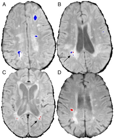
Examples of segmented de novo and repeat lesions. (A) shows large de novo (blue) lesions and (B), a lower slice from the same subject, shows de novo and repeat (red) lesions in close proximity (arrow). (C) shows periventricular repeat lesions with no de novo activity (arrow) and (D) shows a large repeat lesion.