
Figure 2.
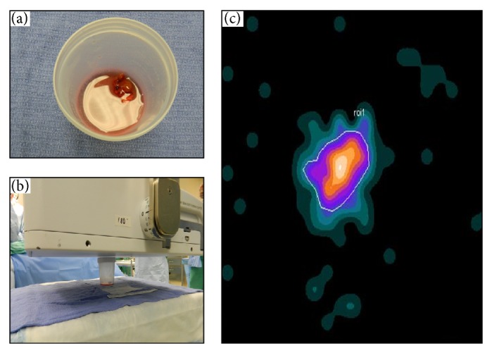
Digital photo of postexcision specimen (a), the intraoperative LFOVGC setup for imaging the specimen (b), and the resultant intraoperative LFOVGC image of a single MIBI-avid specimen, representing abnormal parathyroid tissue (c).
Official websites use .gov
A
.gov website belongs to an official
government organization in the United States.
Secure .gov websites use HTTPS
A lock (
) or https:// means you've safely
connected to the .gov website. Share sensitive
information only on official, secure websites.
Digital photo of postexcision specimen (a), the intraoperative LFOVGC setup for imaging the specimen (b), and the resultant intraoperative LFOVGC image of a single MIBI-avid specimen, representing abnormal parathyroid tissue (c).