Figure 2.

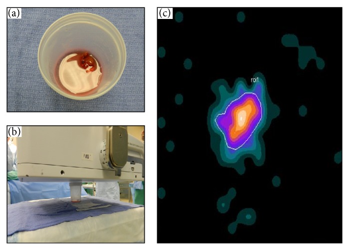
Digital photo of postexcision specimen (a), the intraoperative LFOVGC setup for imaging the specimen (b), and the resultant intraoperative LFOVGC image of a single MIBI-avid specimen, representing abnormal parathyroid tissue (c).

Digital photo of postexcision specimen (a), the intraoperative LFOVGC setup for imaging the specimen (b), and the resultant intraoperative LFOVGC image of a single MIBI-avid specimen, representing abnormal parathyroid tissue (c).