
Figure 3.
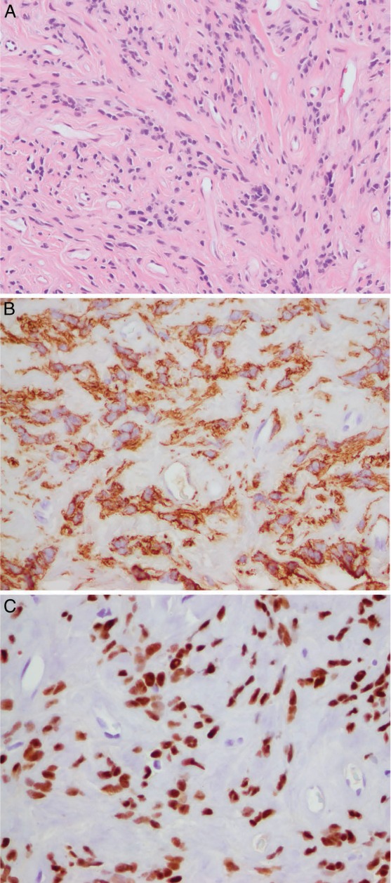
(A) The tumor is composed of bundles of bland spindle cells with “cigar-shaped nuclei,” no nuclear atypia or necrosis, and only rare mitoses (less than 1/10 highpower fields: hematoxylin and eosin stain). (B) The tumor cells were diffusely strongly positive for smooth muscle actin and (C) estrogen receptor and were focally weakly positive for progesterone receptor (not illustrated), which is supportive of uterine origin.