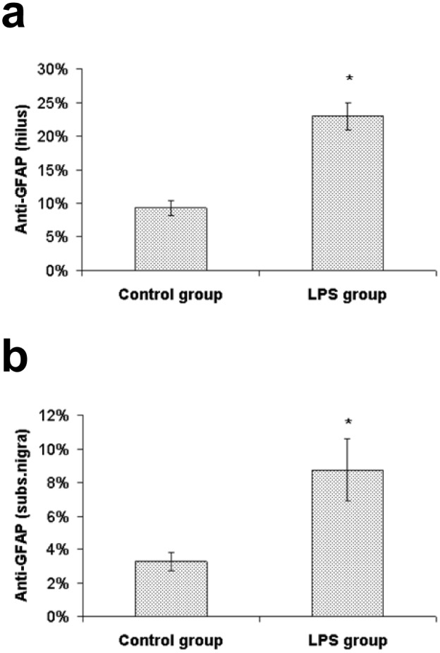
Figure 3. Percentage of the area containing GFAP immunoreactivity.

Values (%) represent mean ± SEM (* indicates p<0.05 from the control group). a) hilus of the dentate gyrus, b) substantia nigra.

Values (%) represent mean ± SEM (* indicates p<0.05 from the control group). a) hilus of the dentate gyrus, b) substantia nigra.