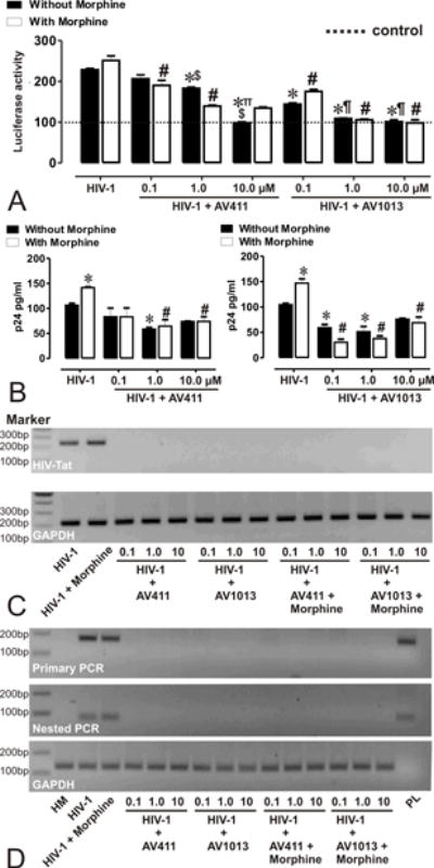
Figure 1. AV411 and AV1013 inhibit HIV-1 SF162 replication in human microglia and displays non-specific interactions with morphine.

(A) HIV-1 Tat protein was monitored by transfecting human microglia with pBlue3’LTR-luc reporter followed by infection with R5 tropic HIV-1 ± morphine ± increasing concentrations of AV411 and AV1013. Relative Tat protein expression was examined 24h post-infection by measuring luciferase activity and the reported values represents the mean luciferase intensity ± SEM from 3 independent experiments (the dotted line represents un-infected control cells). *p<0.05 vs. HIV-1 R5; #p<0.05 vs. R5 + Morphine; ∏p<0.05 vs. 100 nM AV411; $p<0.05 vs. 1 μM AV411; ¶p<0.05 vs. 100 nM AV1013; §p<0.05 vs. 1 μM AV1013. (B) HIV-p24 Gag levels were monitored in human microglia infected with R5 tropic HIV-1 ± morphine ± increasing concentrations of AV411 and AV1013. Cells were infected for 3 days, and then incubated with control media ± morphine ± increasing concentrations of AV411 or AV1013 for another 2 days. Culture supernatants were used to measure p24 levels by ELISA. *p<0.05 vs. HIV-1 R5; #p<0.05 vs. R5 + Morphine. (C-D) HIV-1 Tat mRNA was monitored in human microglia infected with R5 tropic HIV-1 ± morphine ± increasing concentrations of AV411 and AV1013. Tat mRNA expression levels were examined 24 h post-infection by RT-PCR and Nested PCR, and the PCR products were detected using 2% agarose gels stained with ethidium bromide. Marker indicates the position of a 100 base pair (bp) ladder marker. GAPDH served as an input control HM = human microglia; PL= Tat expressing plasmid.
