
Fig. 4.1.
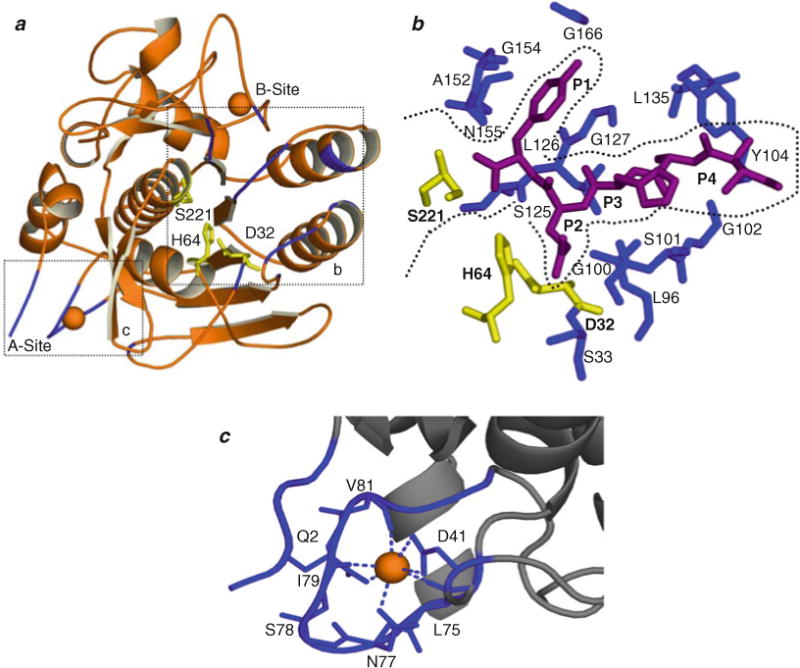
Structural organization of SbtE. (a) Structure of SbtE (ISCJ) is depicted together with the substrate-binding site (b) and the calcium (orange)-binding A-site (c). SbtE also has a second calcium-binding site (B-site) of medium affinity. The catalytic residues are highlighted in yellow. (b) The substrate-binding site is highlighted with an inhibitor (magenta) bound in the S1–S4 pocket. Residues lining the substrate-binding pocket are highlighted in blue. (c) Calcium binding at the A-site is coordinated by residues from a loop comprised of residues 75–83, an N-terminal Gln, and an Asp.