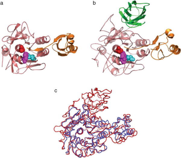
Fig. 4.9.

Structural conservation among IMCs of SbtE, furin, and PC1. All subtilases have a well-conserved subtilisin-like catalytic domain and are stabilized by bound metal ions. Yellow spheres depict calcium ion, while the orange sphere represents sodium ion. (a) Crystal structure of subtilisin E (1SCJ), a bacterial alkaline serine protease. (b) Crystal structure of furin (1P8J), a eukaryotic subtilisin-like protease, with the P-domain and an inhibitor (magenta). (c) Structure of a Ak.1 protease (1DBI), a thermostable subtilase from Bacillus showing the location of several calcium ions.
