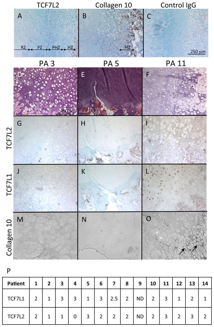
Figure 1.
Immunolocalization of TCF7L2 in exostoses and rib growth plates.
A-O, Sections of rib growth plate obtained from autopsy (A-C) and exostoses obtained from HME patients (PA 1-14) (D-O) were subjected to immunochemical staining forTCF7L2 (A. G-I), collagen 10 (B, M-O), TCF7L1 (J-L) or control IgG (C). D-F are the images of hematoxylin and eosin staining corresponding to G-I, respectively. The bar represents 250 mm for A-O. Note that collagen 10 was only detected in the limited region (O, arrows). P, TCF7L1 and TCF7L2 staining results were scored as 1-3 (0,negative; 1, marginal; 2, positive; and 3, strong positive). ND, not determined.

