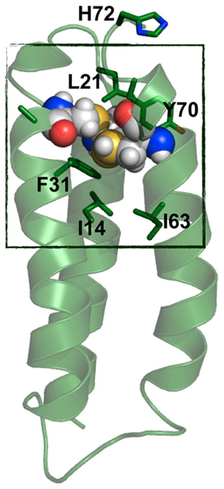
Figure 31.

PyMOL model of α3DIV. Cys mutations are shown in spheres. The box represents the “hydrophobic box”, which includes residues Leu21, Tyr70, Phe31, Ile14, and Ile63. This model is made based on the solution NMR structure of α3D (PDB code: 2A3D751).
