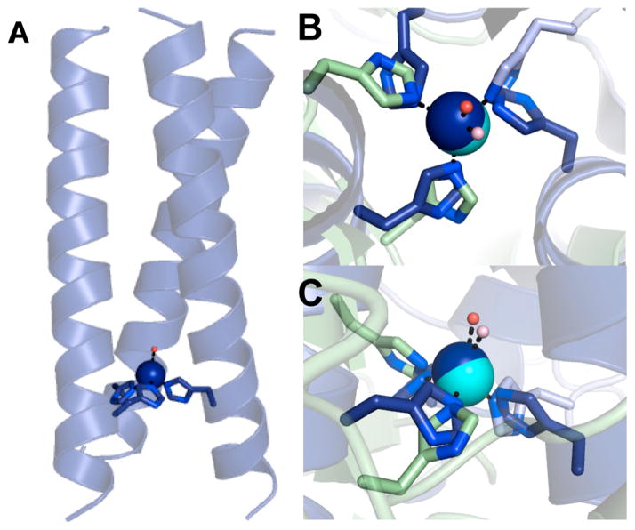
Figure 47.
(A) A model of Cu(TRIL23H)32+/+ based on the crystal structure of Hg(II)SZn(II)N(CSL19PenL23H)3+ (PDB code: 3PBJ213). Overlay of the Zn(II)(His)3(OH2/OH−) site in Hg(II)SZn-(II)N(CSL19PenL23H)3+ (protein ligands: dark blue; zinc: dark blue sphere; zinc-bound water: red sphere) and the T1Cu center in CuNiR from R. sphaeroides (PDB code: 2DY2.958 Protein ligands: light green and teal; copper: cyan sphere; copper-bound water: light pink sphere). (B) Top view. (C) Side view. Adapted with permission from 214. Copyright 2012 National Academy of Sciences.

